
Behavior of lipid droplets could unlock secrets of brain cancer
Biomedical professor receives $433,323 NIH grant to use new technique to see into live cells

Gliomas are among the deadliest kind of brain cancer, causing 80 percent of all malignant tumors. They start in the glial cells and spread quickly to the surrounding tissue and spine.
One aspect about how gliomas form and spread that is not well understood is the increased formation of lipid droplets in the cancer cells. The organelles are essentially packets of fat and oils that play multiple metabolic functions in healthy cells, but it has proven difficult to study them in living specimens.
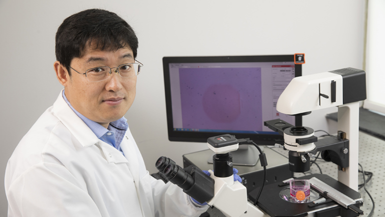
Binghamton University Assistant Professor Fake “Frank” Lu is researching the answer to this problem, using stimulated Raman scattering microscopy. Lu and his team determined the right laser power levels and used the technology to image live cells without staining or other chemical labels, giving the best view yet of what is happening inside cancer cells.
A faculty member at the Thomas J. Watson College of Engineering and Applied Science’s Department of Biomedical Engineering, Lu recently received a three-year, $433,323 grant from the National Institutes of Health to use this method to advance our understanding of the behavior and fate of lipid droplets.
“A cancer cell contains a lot of lipid droplets, and they have been largely ignored in traditional biology,” Lu said. “Chemical fixation and histological staining usually remove the lipid droplets. We have a perfect technology to image these droplets in their fresh, native condition in live cells.”
Lu began his interest in brain tumor study during his post-doctoral research, testing methods for direct imaging and developing clinical technologies. When he arrived at Binghamton three years ago, he focused on studying cancer metabolism and the role that lipid droplets play.
Some droplets are 1 micrometer in size — about 20 times smaller than the width of a human hair — but others can be around 50 nanometers, which is 20 times smaller than a micrometer. A cancer cell tries to store many more droplets than a normal cell, but scientists don’t quite understand why. One theory is that they store nutrients for their metabolism and aggressive growth in harsh tumor environments.
“We know that the brain is a fatty organ, and that cancer cells invade into the normal tissue to break it down,” Lu said. “The glioma cells are very aggressive, and we’re trying to understand why they grow and move so rapidly.”
He theorizes that the lipid droplets may also protect malignant cells from something called lipotoxicity, a metabolic syndrome that accumulates a lethal amount of fatty acid that leads to cellular dysfunction and death. If researchers understand more about the droplets, a cancer treatment could block their synthesis, leading to a buildup of free fatty acids that could kill the cancer cells.
Using stimulated Raman scattering microscopy, Lu also hopes to be able to track the migration of live cancer cells. Cancer cells tend to move away from the tumor core, and there might be a correlation between the lipid contents and the cells’ migration speed and capabilities.
“At the tumor margin, those viable cancer cells contain more lipid droplets,” Lu said. “This was a strong piece of evidence that shows viable cancer cells migrate away from the tumor core, because the core is too difficult of an environment for cell survival. These lipid droplets are helping their migration in some way — that’s our hypothesis, at least.”
Lu conducted preliminary experiments on mice that helped to persuade the NIH grant panel to fund wider study. Also attractive to the NIH was Lu’s plan to train and pay nine undergraduates to conduct research. Their tasks will include treating and imaging the live cells with different types of fatty acids to investigate their effectiveness in curbing cancer growth.
“This project has small and handy experiments using the same imaging setup that can expose undergraduate student researchers to hands-on biophotonics research,” he said. “This is a strength that the grant reviewers really loved, and I do, too.”
Lu’s research project, “Low-Power Stimulated Raman Time-Lapse Microscope for Tracking Dynamics and Fate of Lipid Droplets in Glioma Cells,” is funded through an R15 grant from the National Institute of General Medical Sciences.