A look inside: MRI partnership gives lab students a chance to study a living brain
A partnership between Binghamton University and UHS aids academic and clinical research, in addition to meeting patient needs

Over their professor’s shoulder, the neuroscience students peered inside a man’s skull.
SUNY Distinguished Professor of Psychology J. David Jentsch took the class layer by layer, shifting effortlessly through structures and ventricles in ghostly gray, some lit with specks of light to indicate activity.
“Did you know that the cerebellum has as many neurons as all the rest of the nervous system?” he quizzed.
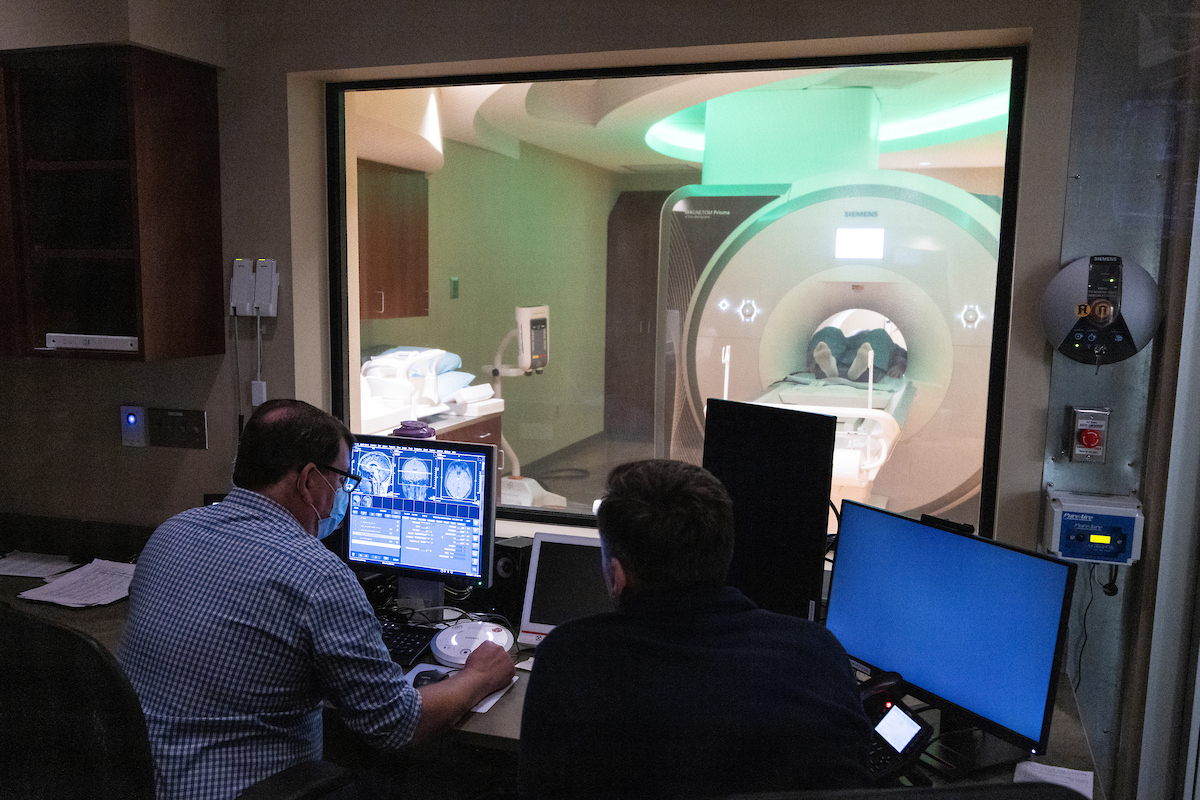
The detailed glimpse into a living brain came courtesy of a state-of-the-art magnetic resonance imaging (MRI) scanner located across the street from campus at United Health Services’ Vestal facility. The result of a partnership between UHS and the University, the $2.6 million Siemens Magnetom Prisma 3 Tesla scanner is used for academic and clinical research, as well as the region’s medical needs.
Jentsch, who leads the University’s Brain and Body Imaging Research Center, took small groups of students from his neuroscience laboratory course to UHS in October, giving them the opportunity to observe how MRI scans are conducted and evaluated. The “patients” were volunteers, and no medical information was disclosed.
The machine weighs 14 tons, largely due to the vast coil of wire it needs to create a magnetic field 60,000 times stronger than that of the Earth. In order for the magnet to act as a superconductor, the coil is bathed in 1,000 liters of liquid helium.
“Today, the only way we know how to achieve superconduction is at very low temperatures, lower than minus 400 Fahrenheit,” Jentsch explained. “Because of that, there’s zero resistance in the wire. And that current runs forever, unless we release the helium and bring it back up to temperature.”
In other words, the current can never be turned off — even if the building were to lose power.
Before he entered the room, the research subject was scanned for any metal on his body — an important consideration, because the powerful field would forcefully pull any ferromagnetic item into the machine. Steel and copper bars within the room’s walls keep the field from spilling outward and disrupting the technology in adjoining rooms.
The subject enters the machine head first, wearing a plastic mask that improves the signal-to-noise ratio, allowing for improved brain imaging. As it generates images, the scanner produces around 100 to 125 decibels of noise, akin to a jumbo jet; in addition to having no metal on their person, everyone in the room requires headphones, which also allow the technologists to communicate with the test subject.
First, the technician created a localizer: a low-resolution picture that helps determine whether the position of the subject’s head is correct. In the scans that followed, the MRI took a series of pictures of the brain, averaging them together for the cleanest possible image.
Essentially, the magnetic field is so strong that the protons in the body align with it. Jentsch explained that the proton density of different bodily tissues is distinct, allowing for the creation of layered images.
Water-filled cavities in the brain and body are dark in the images because they have a greater density of protons than tissue. The scanner is sensitive enough to detect the movement of fluid in tissue and along axons in the brain’s white matter, using color coding to indicate the movement’s direction. This allows technologists to see, for example, the progressive loss of brain organization with diseases that affect the white matter.
During the sessions, Jentsch assigned the test subject a task: to think up words starting with a particular letter, but silently, to keep from firing the brain’s motor cortex. Reviewing the resulting scan with students, Jentsch pointed out a lit area that showed activity: known as the Broca’s area, it’s central to language.
While useful for research, tests like this one are also used in medical procedures, Jentsch explained. Surgery to treat severe epilepsy, for example, may involve removing afflicted portions of the brain; technologists would identify the location of the brain’s speech centers before the procedure to make sure they remain untouched.
While the scans were being set up, students chatted about their own experiences with MRIs. Several had imaging done, they said, but the machines weren’t as high-tech as this one.
“It’s definitely something I didn’t think I would experience in class,” reflected Edward Almonte, a senior neuroscience major on the pre-healthcare track. “We’re very fortunate to see this operated.”