


Innovation could transform brain surgery
Surgical removal of brain tumors may become easier and more precise, thanks to Binghamton University research that recently received funding from the National Institutes of Health.
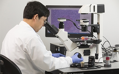
Fake “Frank” Lu, an assistant professor of biomedical engineering, uses stimulated Raman scattering (SRS), a molecule identification technique, to develop a multicolor imaging technology for brain cancer pathology during surgery.
A three-year, $750,000 R00 grant from the National Institute of Biomedical Imaging and Bioengineering (NIBIB) supports his research at Binghamton.
There are two steps to get rid of a brain tumor: removing the literal mass and then removing the lingering cancer cells at the edges. Removing the lingering cells can be tough, requiring evaluation to avoid destroying functioning brain structures.
Cancer cells are denser than normal cells, which helps them to be identified. Modern procedures involve a pathologist standing by for intraoperative consultation, using neuronavigation systems (such as an MRI scan) or fluorescence imaging to detect lingering cancer cells. Each of these techniques has drawbacks:
With intraoperative staining-based histopathology, tissue samples are taken and evaluated in a nearby laboratory. Although accurate, this is ultimately a slow process, only allowing three samples to be evaluated during surgery.
Neuronavigation systems can also be flawed as a result of the brain moving during the surgery, which is called “brain shift.”
Fluorescence imaging uses fluorescent dyes to label and locate the cancer cells. Unfortunately, this is a messy process and isn’t accurate at distinguishing the margins of the tumor.
Lu’s technology is label-free, rapid and detailed. He expects that in the future, SRS can be used to evaluate 20-30 tissue samples during surgery to help delineate the tumor margin.
SRS detects “molecular fingerprints” by exciting the chemical bonds in molecules and reading the frequencies emitted by the vibrational states of the bond. This information is then used to assign different colors to the molecules, allowing for a detailed image.
Lu has also improved SRS so that it can create images of lipids, fatty acids that he says are important markers for the presence of cancer cells. Axons, the long, thread-like sections of nerve cells, are wrapped in lipid layers called myelin sheath. In areas affected by cancer, they break down, leaving lipid droplets that can be easily detected by Lu’s technology.
Lu envisions the technology being incorporated into a machine for operating rooms.
Read more about his work in Discover-e.