Mass Spectrometers
Q Exactive™ HF Hybrid Quadrupole-Orbitrap™ Mass Spectrometer: State-of-the-art proteomics
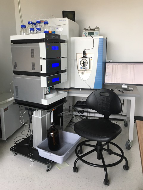
Equipped with the most recent, state-of-the-art instruments including the UltiMate™ 3000 nano LC System connected to a Q Exactive™ HF Hybrid Quadrupole-Orbitrap™ Mass Spectrometer, the laboratory is also equipped with necessary labware for sample preparation, including one- and two-dimensional gel electrophoresis for protein separation, a high-resolution densitometer for gel and western blots imaging, an offline HPLC for peptide and protein separation and the necessary software packages for protein identification and quantification.
Additional equipment includes a Meso Scale Discovery U-Plex ELISA platform for multiplexing ELISA assays suitable for biomarkers and protein targets validation.
Standardized techniques include:
- High throughput LC-MS/MS analysis for protein identification/quantification using both stable isotope labeling strategy (TMT, SILAC, 15N labeling) and label free strategy.
- Characterization of post-translational modifications of proteins (phosphorylation, glycosylation)
- Targeted protein quantification in complex biological and clinical samples
- Biomarker studies
- Development of pharmacodynamic biomarkers in pre-clinical and clinical studies
The proteomics laboratory encourages research collaborations across campus, nationally and internationally.
The School of Pharmacy thanks the Decker Foundation for its continuous support and the generous gift to cover the cost of our state-of-the-art instruments.
Waters Autopurification System with SQD2 MS and PDA detectors
This multi-purpose instrument is used for purification of pharmaceutical agents and for the routine analysis of synthetic reactions. It is equipped with an SQD2 (single quadrupole) mass spectrometer with electrospray ionization capable of detecting compounds up to 3000 m/z. The pumps on this instrument are capable of delivering from 0.1 mL/min (analytical scale chromatography) to 150 mL/min (prep scale chromatography). The quad pumps can select from among four buffers, thus allowing facile switching between formic acid buffers (for analytical work) and TFA buffers (for preparative work). A column switcher can select from among five columns. The system is equipped with MaxEnt 3.1 deconvolution software, allowing for the analysis of protein samples, including antibodies. Our lab routinely uses this instrument both for purification/analysis of organic compounds and for studying antibody modifications.
Waters Acquity UPLC (H-class) with QDa detector
Used for the analysis and quantitation of dilute mixtures of pharmaceutical and biopharmaceutical preparations, this instrument is equipped with a dual-wavelength detector, a fluorescence detector and a single quadrupole mass detector (QDa), thus allowing for a wide range of detection capabilities. The narrow bore tubing and high pressures used in this system (up to 15,000 psi) allow for very fast separations and very high sensitivity (~50 pg). A switching valve allows selection from among four columns and four buffers. Open access software allows users to easily select an appropriate method and log samples into a queue. Auto-processing methods then generate a PDF that can be directly attached into notebooks. This system is routinely used for reaction monitoring, kinetic studies and pharmaceutical stability studies. Both protein and small molecule methods are available.
Waters Xevo TQD Mass Spectrometer
The Waters Xevo TQD is a tandem quadrupole LCMS connected to an H-class UPLC. This
instrument is particularly useful for: 1) drug/metabolite detection in biological
matrix; 2) Environmental analysis of trace organic compounds; and 3) Structural determination
of organic/natural products. Tandem quadrupole LCMS using MRM analysis is typically
considered the "industry standard" method for small molecule PK analysis and metabolite
monitoring during in vivo studies. In addition, it is suitable for protein/peptide
quantitation using a protease digestion approach. The LOD for this instrument varies
considerably between samples, but routine analysis of common drugs usually gives an
LOD of 5-50 picograms with minimal optimization.
Nuclear Magnetic Resonance (NMR) Spectroscopy
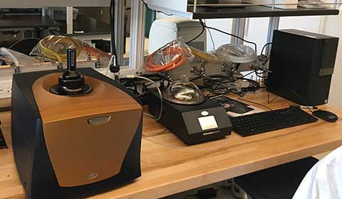
Bruker Avance III HD 400
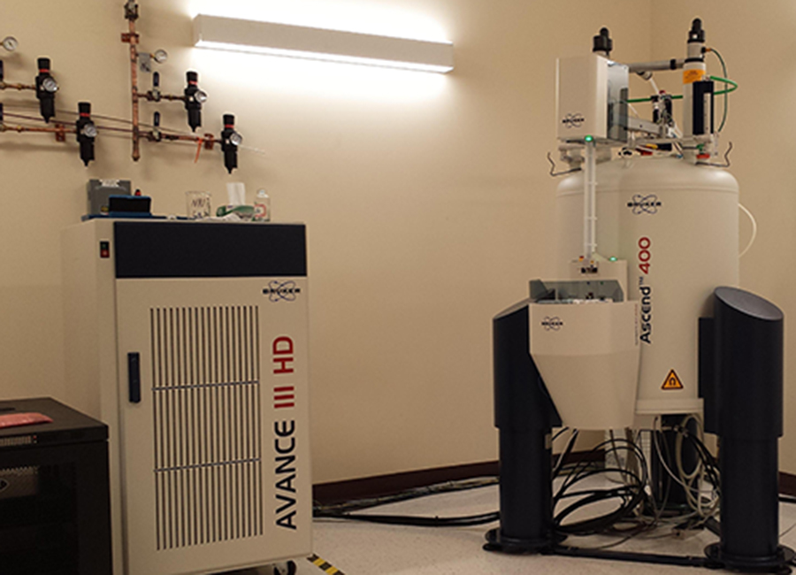
The Bruker Avance III HD 400 contains a 9.1 Tesla shielded, superconducting magnet and performs proton NMR analysis at 400 MHz. It is connected to a 24-sample automated carousel. The primary probe is a 5 mm Broadband/Fluorine probe that is typically used for structural analysis of organic and inorganic compounds in solution. It is equipped for pulsed field gradients and can be tuned automatically to detect almost all nuclei. Typical experiments performed with this probe include all two-dimensional experiments, i.e. COSY, NOESY, HSQC, HMBC and many more.
The School of Pharmacy thanks the Decker Foundation for its continuous support and the generous gift to cover the cost of our state-of-the-art instruments.
PCR Detection Systems
CFX96 and CFX384 Real Time PCR Detection Systems
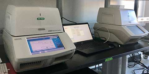
These are real-time polymerase chain-reaction detection systems that can accommodate 96- and 384-well plates, utilizing five colors and one FRET channel detection to enable single- or multi-plex (up to five target) reactions to determine gene expression. They can run regular or fast PCR cycles using Taqman or SYBR technologies and their automated software can run all analyses for complex data sets. Additional utilities of these systems include any combination of fluorescence and temperature measurements, including FRET Melt and genomic mutation detection. Go online for a video tour and more details on the CFX96 and the CFX384.
Flow Cytometry
BD Biosciences Accuri C6 Plus Flow Cytometer
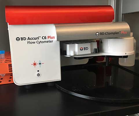
This newest generation of the BD Accuri™ platform is equipped with a blue and red laser, two light scatter detectors and four fluorescence detectors with optical filters optimized for the detection of many popular fluorochromes, including FITC, PE, PerCP-Cy™5.5 and APC, as well as newer polymer dyes such as BD Horizon Brilliant™ Blue 515. It can also analyze many variants of fluorescent proteins, such as GFP, YFP and mCherry. A non-pressurized, peristaltic pump system drives the fluidics. The system accurately monitors the sample volume pulled per run, and can calculate absolute counts or sample concentration per µL without the use of counting beads. Our laboratory is currently using the BD Accuri C6 flowcytometer to monitor cell surface expression of immunological markers on skeletal muscle cells.
Chromatography
Bio-Rad NGC chromatography system
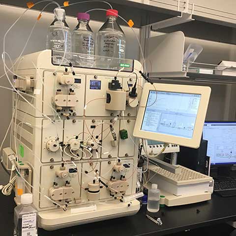
This versatile protein purification system is capable of selecting among eight columns and nine buffers. A sample pump allows for the loading of large volumes of samples (for example, cell lysate for affinity capture chromatography). Detectors include a quad-wavelength UV detector, pH sensor and conductivity detector. A BioFrac system collects fractions into either tubes or 96-well plates. Methods are currently set up for size-exclusion and hydrophobic-interaction chromatography. This system is also ideally suited for ion exchange and affinity chromatography methods.
Spectrometry
Jasco J-1500 Circular Dichroism Spectrophotometer
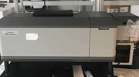
This spectrophotometer allows for the simultaneous measurement of circular and linear dichroism (CD and LD), as well as absorbance, for the determination of achirality in proteins, nucleic acids and chemical moieties. Our J-1500 is outfitted with a dedicated stopped-flow system for kinetic and protein dynamic studies, and with a peltier unit for temperature control, enabling studies of achirality as a function of temperature (e.g. melting studies). Spectra Manager software functions for both control and data analysis purposes. Go online for more details.
Agilent Cary 60 UV-Vis Spectrophotometer
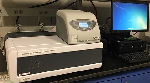
Efficient, accurate and flexible, this spectrophotometer maintains high sensibility with micro volume samples. The unique Agilent Xenon flash lamp source, along with the optical configuration of the instrument, eliminates photodegradation, and thus, the measurements are precise and reproducible.
Nanodrop 2000 by Thermo Fisher
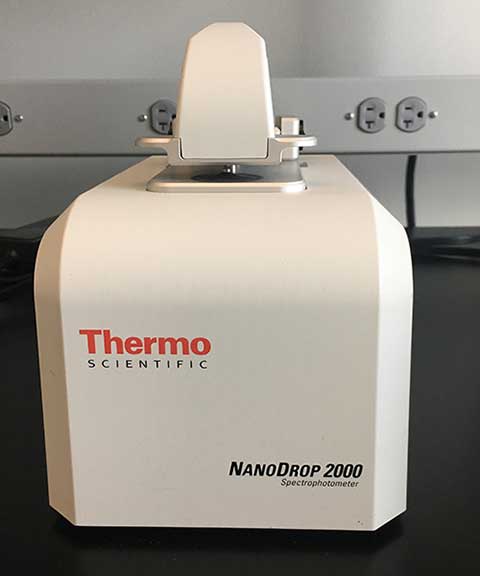
This spectrophotometer is capable of determining the concentration of DNA, RNA and proteins from only 1-2 L of sample. It can measure absorbances in the full UV-Vis range with a wide range of applications, including determining the optical density of e. coli solutions, the concentration of a drug with absorbance in the UV-Vis range against a standard curve and more. Visit the web for more details and to view a short video.
Molecular Devices i3x Microplate Reader
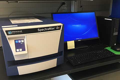
The Molecular Devices SpectraMax i3x Multi-Mode microplate reader measures spectral-based absorbance, fluorescence and luminescence. The SpectraMax Injector Cartridge with SmartInject™ Technology for the SpectraMax® i3x Multi-Mode Microplate Detection Platform supports flash-based applications and includes dual luciferase and ATP assays. The SpectraMax i3x’s Spectral Fusion™ illumination allows for increased sensitivity across entire excitation ranges. The photomultiplier tube (PMT) is cooled to allow for better detection in very low light applications. User-exchangeable modular upgrades are also available from the manufacturer. These include modules for Western Blot Detection, Imaging, Fast Kinetics with Injectors, Time-Resolved Fluorescence and HTRF.
Gel Scanners and Gel BioMolecular Imaging
Amersham Typhoon 5 BioMolecular Imager
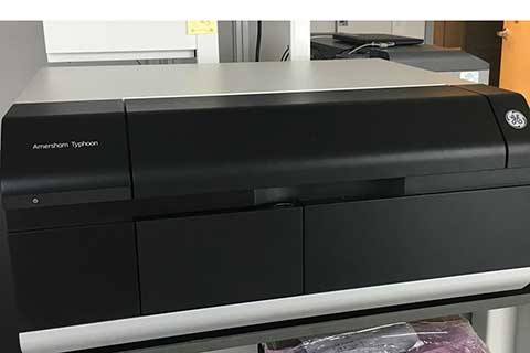
This laser scanning platform enables imaging of electrophoretic gels and membranes in near-infrared and RGB fluorescent ranges, as well as through phosphor imaging or colorimetrically stained samples, such as through Coomassie blue or silver stains. The scanning area can accommodate materials up to 40 x 46 cm, and the scanned pixel size ranges from 10-200 m (or a 1000 M pre-scan). Fluorescent filters available include IP (390BP), Cy2 (525BP20), Cy3 (570BP20), Cy5 (670BP30), IRshort (720BP20) and IRlong (825BP30); phosphor imaging allows for detection of energy produced from common isotopes, such as 3H, 11C, 14C, 125I, 18F, 32P, 33P, 39S and 99mTc. Common uses include large polyacrylamide DNA sequencing gel imaging, western blots using NIR-tagged secondary antibodies, autoradiographs from physiological samples and much more. See the website with a short video for more details.
Azure c300 Gel Scanner
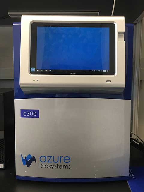
This digital darkroom enables detection and quantification of a variety of electrophoretic assays. It can be used for applications that require trans- or epi-white light exposure, epi-blue or trans-UV (302 and 365 nm) light and chemiluminescent detection, including PCR and DNA agarose gels, polyacrylamide gels and western blots. The field of view has a maximal area of 20 x 15 cm and images are obtained with an 8.3 MP cooled camera. Go to the web for more details.
Imaging
Leica DMi8 CEL Advanced Confocal Microscope
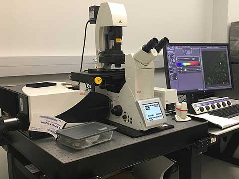
The Leica DMi8CEL Advanced confocal microscope is equipped for confocal imaging of mounted slides or live cell cultures with an inverted scanning stage. The system uses a scan optics module VISIR with rotation and SP8 field of view scanner. Two internal detector channels (PMT) and two internal detector channels (HyD) allow multicolor imaging with the laser kit RYB for AOBS and a 405 nm DMOD flexible laser. The True Confocal Scanning (TCS) system provides planar 1–1800 Hz imaging with a maximum resolution of 64 Mpixels. Objectives allow magnification at 10X, 20X glycerin immersion, 40X oil and 63X oil. An epifluorescent lamp is available for initial sample viewing (DAPI, FITC, DsRed). Live cell images can be collected with the live cell chamber to maintain temperature, humidity and CO2 using 35mm dishes with a #1.5 coverglass bottom.
The School of Pharmacy thanks the Decker Foundation for its continuous support and the generous gift to cover the cost of our state-of-the-art instruments.
Olympus VS120-S6-W slide loader system
This system allows for manually loading from one to six standard slides, along with any associated meta-data. The VS120 not only creates high resolution brightfield images, but can also scan in full multi-fluorescence mode with the X-CITE 120LED Boost lamp. The system is designed for imaging the following fluorophores: DAPI (blue), FITC (green), TRITC (orange/red), MCherry (red) and Cy5 (far red). Utilizing virtual microscopy for fluorescence imaging helps to minimize problems associated with damaging and fading of sensitive fluorescence samples. An innovative new algorithm makes image stitching more precise than ever, enabling high-level accuracy that can be applied from small animal brain slices to large specimens. The VS120 also switches seamlessly between micro- and macro-observation to enable swift viewing of regions of interest and overall structures alike. UPLSAPO 2x, 4X, 10x, 20x and 40x objectives are available, allowing users to choose the objective most suitable for their research needs. Automatic specimen recognition capability limits scanning to the specimen area, with high-level color fidelity and image quality. Images are acquired with the VS-HIGHSENS V2.9 software and can be analyzed with VS-ANALYSIS V2.9.
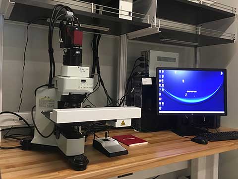
Olympus IX73 inverted microscope
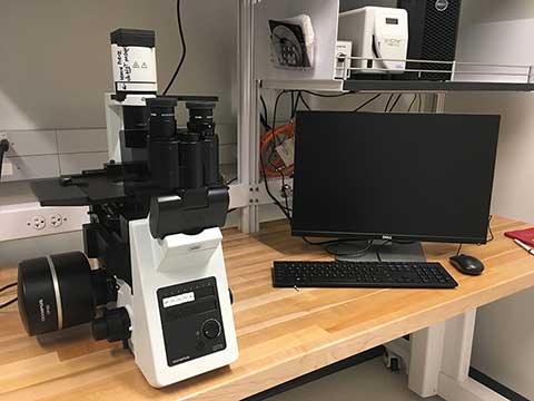
This is a white light and epifluorescent microscope designed for maximum flexibility. Phase contrast objectives allow for imaging live cells through standard tissue culture plastic dishes or fixed slides. The microscope is entirely manual in operation with 4X, 10X, 20X and 40X objectives. The system is designed for imaging of fluorophore wavelengths of BFP (blue fluorescent protein), FITC (green), CY3.5 (mStrawberry) and CY5. High-quality images can be captured with the DP80 dual color-monochrome camera and Olympus CellSens software.
Olympus CKX53SF-1-2 Microscope
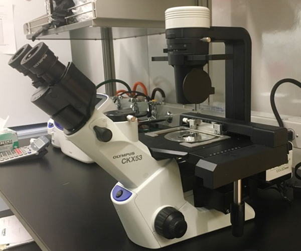
The Olympus CKX53 inverted microscope is specially developed for cell culture applications with a small footprint and lightweight design. The microscope is paired with a long-life X-Cite 120LED lamp and features integrated Phase Contrast (iPC) quality for improved brightfield imaging. Inversion contrast technology improves clarity of three-dimensional views and enables observation of the bottom two layers of a multi-layer flask with the 4X objective. Epifluorescent imaging is available for blue, green and red fluorophores and the "Umbra shield" enhances fluorescent contrast and allows imaging even in bright-light room conditions. Magnification is available with 4X, 10X and 20X objective. The system can be upgraded with a camera for image collection, but no camera is currently attached.
In-Vivo Equipment
Perkin Elmer IVIS Lumina LT Series III Imaging System
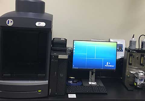
This system allows for rapid, non-invasive in-vivo imaging using fluorescence and bioluminescence. It can also accommodate Petri dishes or micro-titer plates for in-vitro imaging. This system is equipped with filters that can be used to image reporters that emit from green to near-infrared. The imaging system is equipped with an adjustable field of view from 5-12.5 cm, with a field of view that can expand up to 24 cm, allowing for imaging of up to five mice or two medium-size rats. The IVIS Lumina LT CCD camera is 13 x 13 mm square, with 1024 x 1024 pixels 13 micron in width, which yields higher imaging resolution. The imaging chamber is equipped with a heated stage to maintain optimum body temperature for animals. The IVIS Lumina also has an integrated gas anesthesia component, with five gas anesthesia ports and five position manifolds within the imaging chamber to allow for anesthesia maintenance throughout the imaging sessions. The system also has a Living Image Software set up to the computer to allow for simple analysis for the user in both fluorescent and bioluminescent modes. Our laboratory is planning on using the IVIS Lumina system to assess skeletal muscle inflammation in live mice by cathepsin activity using Prosense 680.
ECGenie
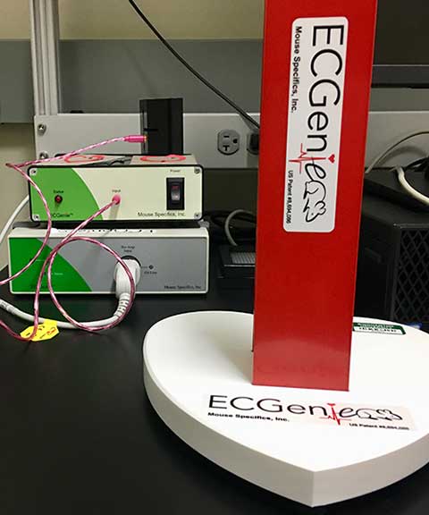
This provides a rapid, non-invasive solution for recording electrocardiograms (ECGs) in awake laboratory animals. Companion EzCG analyses software analyzes the signals to assess animal health, cardiac diseases and drug toxicity. Applications include arrhythmia detection, health monitoring and drug screening in fragile transgenic and knockout animals, including newborn pups. We use this system to determine if a particular drug or genetic mutation causes disturbances in heart rate variability and/or cardiac electrical conduction responses. We can also show whether therapeutic interventions are capable of reversing abnormal ECGs.
Mikro-Tip® Pressure Volume System (MPVS) Ultra Foundation System
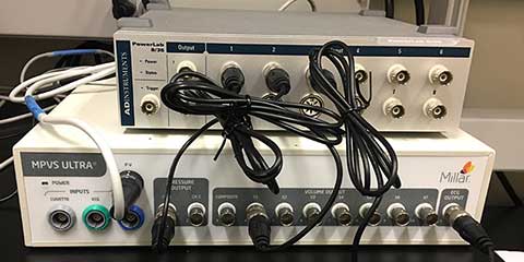
This equipment is used to determine cardiac function in live animals. Specifically, this system measures left ventricular pressure and volume, commonly referred to as PV loops. Data is acquired through PowerLab hardware, then subsequently processed and analyzed using LabChart software. We use this system to determine if a particular drug or genetic mutation causes cardiac dysfunction and can decipher what kind of dysfunction is occurring. Subsequently, this system is also used to reveal if therapeutic interventions are capable of normalizing cardiac function
Detection Systems and Bioanalyzers
The MSD Meso QuickPlex SQ120
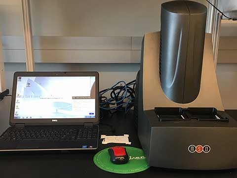
This is an electrochemiluminscence (ECL)-based detection system for single or multiplex immunoassays that offers simultaneous analysis of up to 10 analytes per well in a 96-well plate format, with detection of analytes at levels as low as 0.05 pg/mL. A variety of assays are available directly from MSD including panels to measure markers indicative of kidney injury, cardiac damage, and inflammation in human and animal models. Additionally, custom assays may be developed with user-supplied antibodies, aptamers or other biological recognition elements. We have been using this platform for cytokine profiling and biomarker assessments in response to drug treatment or disease state.
Agilent 2100 Electrophoresis Bioanalyzer
This bioanalyzer uses a micro-capillary based electrophoretic cell that allows rapid and sensitive investigation of nucleic acid samples. It is the gold standard for sample quality assessment of nucleic acids, including total RNA, labeled RNA, small- and micro-RNAs as well as small and large DNA fragments. The bioanalyzer provides precise information on fragment size distribution, but is not recommended for accurate quantification. Users are expected to provide their own cassettes and reagents.
Miscellaneous Equipment
BioRad Protean i12 IEF System with Criterion Cell
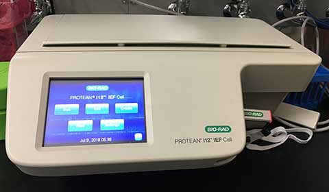
This equipment is suitable for high-resolution, two-dimensional separation of complex protein mixtures in biological and/or clinical samples. Separated proteins can then be identified by mass spectrometry and their relative abundance, determined from their spot density in the 2-D gel image. The technique is commonly used to identify proteins that are altered in their levels between different samples and also to characterize post-translational modifications of proteins of interest.
Nano ITC Calorimeter
This low-volume isothermal titration calorimeter is designed to provide maximum sensitivity and flexibility for the label-free study of biomolecular binding. Affinity, enthalp, and stoichiometry information can be generated from the measurement of heat effects as small as 100 nanojoules.
Biotek Instruments MultiFlow FX Microplate Dispenser
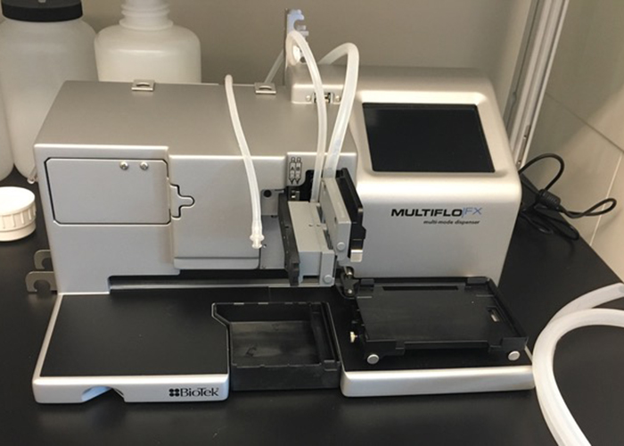
The Biotek Instruments MultiFlow FX Microplate Dispenser is a programmable plate washer that is used in concert with our Meso QuickPlex SQ 120 and SpectraMax i3x plate reader for facilitating wash steps in heterogeneous microplate assays. It also offers magnetic bead handling and adherent cell-washing capabilities.