Energy Consumption of Cell Migration
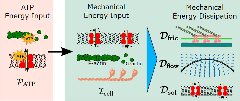
We apply physics relations that satisfy energy identities to quantify energy consumption during cell migration under different physical environments. The results show that the cell energy consumption is higher when cells migrate in viscous or narrow environments, conditions that cancer cell metastasis happens. The work adds new information to the coupling of metabolic processes with other cell biophysical behaviors.
Higher Hydraulic Resistance Accelerates Water-Driven Cell Migration
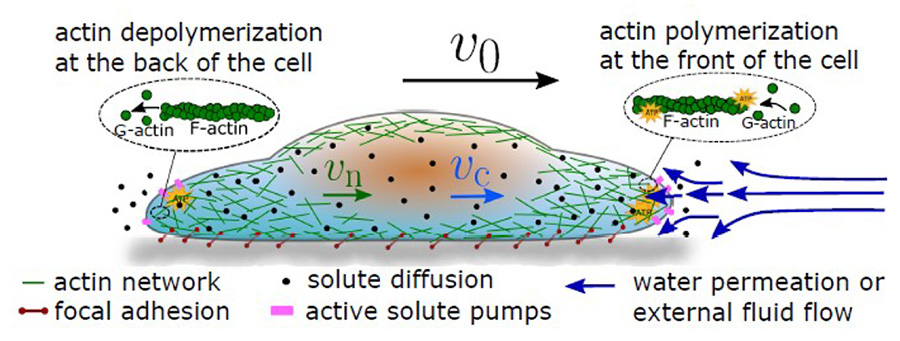
Cells in vivo live in diverse physical environments that provide mechanical cues for cells to deform, migrate, and carry out their biological functions. In the two phas emodel, actin network and cytosol are modeled as two fluids. The actin network forms focal adhesions with the environment. Solutes diffuse within the cell and are transported across the cell membrane. Water flux happens across the cell membrane when the chemical potential of water is imbalanced. Actin polymerization and depolymerization happen at the front and back of the cell, respectively.
Cells Can Migrate under External Electric Fields by Ion and Water Transportation
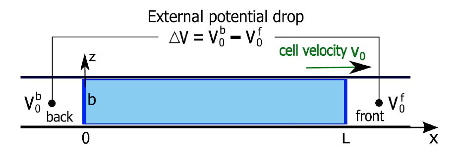
A diagram of a 1D cell model in an applied external voltage drop, ΔV. V0b and V0f are the prescribed extracellular electric potential at the back and front of the cell, respectively.
Ionic Contents in the Medium Regulate Cell Volume at the Single-Cell Level
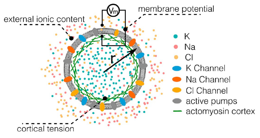
Cell volume regulation on short timescales is closely related to ionic regulation. Major species in the cell are Na+, K+, Cl−, and impermeable charged proteins. Ion concentrations are controlled by passive ion channels and active ion pumps. Changes in ionic content also influence the transmembrane potential. The cell volume, which is determined by the overall water and protein content, can be modulated by changes in the transmembrane potential, external ionic content, or cortical tension.
Water Permeation Can Contribute to Cell Division
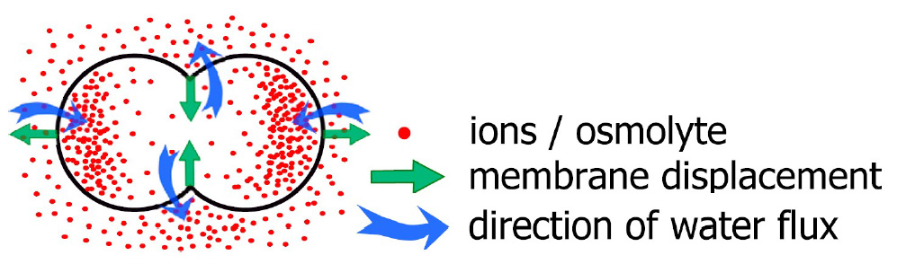
A possible spatial arrangement of intracellular ionic sistribution during cell division. The cell shape is determined by the water flux across the membrane. The water flux is determined by osmotic and hydrostatic pressure differences across the cell membrane.